Thoracic Outlet Syndrome (TOS) is the compression of neurovascular structures as they pass through the cervicothoracobrachial region. The term ‘TOS’ is a generic term that does not specifically identify the structure being compressed. There are two categories of TOS: vascular (VTOS) and neurological (NTOS). However, NTOS is much more common, making up approximately 95-98% of all TOS cases. A combination of both categories of TOS is also possible.

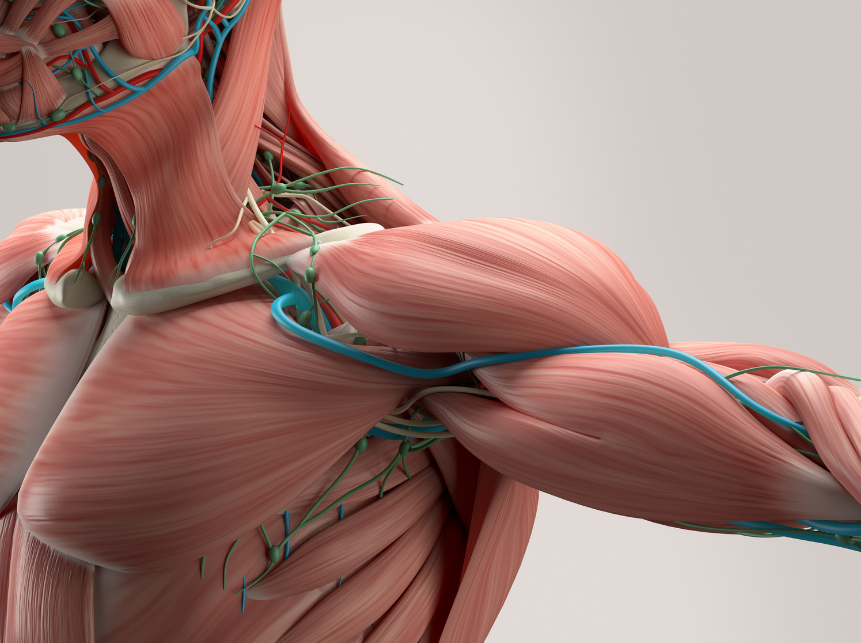
There are multiple locations where compression of structures can occur. One location is a passageway bordered by the anterior and middle scalene muscles (neck muscle) and first rib. These structures can compress on the brachial plexus (neural structures for the upper limb) and the subclavian artery.
A second passageway called the costoclavicular triangle is also a possible compression site for structures. The passageway is formed by the middle third of the clavicle (collar bone), the first rib and the upper border of the scapula (shoulder blade). The subclavian vein and brachial plexus pass through this region. Compression on these structures can also occur as a result of congenital abnormalities, trauma to the first rib or clavicle, and structural changes in the musculature.
The third and final passageway consists of the coracoid process of the scapula (bony protrusion of the shoulder blade), pectoralis minor (chest muscle) and ribs 2-4. Tightening of the pectoralis minor is often a major factor in compression here during shoulder abduction.

Incidence:
TOS is relatively uncommon, affecting approximately 8% of the population. However, TOS is 3-4 times more frequent in women between the ages of 20 and 50 years. The presence of congenital anomalies such as cervical ribs (which are present in approximately 0.5% of the population) can result in symptomatic TOS, however it is more commonly associated with tight fibromuscular structures. Other factors could implicate TOS including postural behaviours, repetitive stress injuries, trauma to the ribs or clavicle, hypertrophy (increased activity) of the scalene muscles, weakness of the trapezii, levator scapulae, rhomboid muscles and pectoral muscles.
Symptoms:
Symptoms often vary from patient to patient due to the multiple areas and structures that can be compressed. Symptoms can vary in intensity from mild pain and sensory changes to limb-threatening complications. Patients can present with pain anywhere from the neck and face, chest, shoulder or upper limb, as well as paresthesia (abnormal sensations such as pins and needles and numbness). Other possible complaints can include weakness and fatigue of musculature, a feeling of heaviness of the upper limb and discoloration/changes of temperature in the skin. These symptoms are typically aggravated with overhead shoulder movements combined with external rotation of the glenohumeral joint (shoulder joint), for example when serving in tennis or painting a ceiling.

What your physiotherapist can do for you:
Conservative management from a Physiotherapist should be the initial strategy in treating TOS. Conservative management under the guidance of a Physiotherapist has been proven to be effective in decreasing symptoms, improving function and enabling return to work/activity. Physiotherapy management consists of patient education, pain control through manual therapies, restoration of joint range of motion, neurodynamic gliding techniques, as well as strengthening and stretching exercises.
For further information regarding Thoracic Outlet Syndrome please contact your physiotherapist!
- Laulan J, Fouquet B, Rodaix C, Jauffret P, Roquelaure Y, Descatha A. Thoracic outlet syndrome: definition, aetiological factors, diagnosis, management and occupational impact. Journal of occupational rehabilitation. 2011 Sep 1;21(3):366-73.


